
By robert a hannaman
Read or Download MedStudy 12th Edition Internal Medicine Board Review Core Curriculum 2007 2008: Nephrology PDF
Similar medicine books
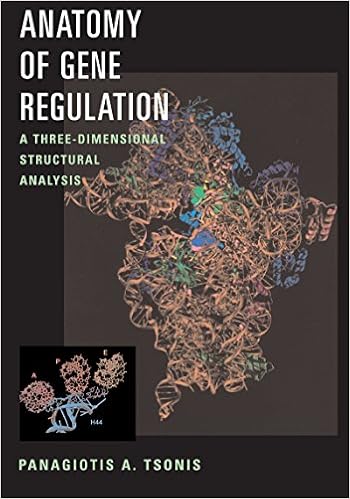
Panagiotis A. Tsonis's Anatomy of Gene Regulation: A Three-dimensional Structural PDF
Not basic line drawings on a web page, molecular buildings can now be considered in full-figured glory, usually in colour or even with interactive chances. Anatomy of Gene legislation is the 1st e-book to offer the components and tactics of gene rules on the three-d point. bright buildings of nucleic acids and their spouse proteins are published in full-color, three-d shape.
- Ayurveda For Dummies
- ABC of COPD
- Upper Gastrointestinal Surgery
- Performing Arts Medicine in Clinical Practice
Extra resources for MedStudy 12th Edition Internal Medicine Board Review Core Curriculum 2007 2008: Nephrology
Example text
Concentrated urine andlor excretion of excessive amounts of stoneforming products cause precipitation and stone formation . Finally, certain products may act as a nidus for stone formation. Calcium stone inducers: hypercalciuria, uric acid, hypocitraturia, hyperoxaluria, and medullary sponge disease. Citrate chelate calcium, thereby preventing stones. Acidosi. ) and also leaches calcium from the bones, resulting in hypercaIciuria. Although the calcium stones are usually a combination, they are often grouped into calcium pho phate and calcium oxalate stones.
Chemistry panel. B. 24-hour urine protein and creatinine clearance. C. ANA. D. Serum complement levels. E. VORL. F. HIV screening. F. HBsAg. G. Renal biopsy. [G . All of the tests should be performed except a renal biopsy. If indicated by these initia l tests. a renal biopsy is done. ] 2 Ie) All the above lab tests (except renal biopsy) are normal. What is the most likely diagnosis? A. B. C. D. E. F. G. Post-infectious GN (PIGN). IgA nephropathy. RPGN. Membranoprolifcrative GN. Minimal change disease.
Affects the liver and pancreas also. Affects the basement membrane, cochlea, and lens. Normal urinary sediment. Persistent microscopic hematuria, which worsens after in fections. Gross hematuria. [I (A) 2 (B. ) 3 (B) 4 (B) 5 (B) 6 (A) 7 (C) 8 (A) 9 (B)] 17) Renal stones: A . Calcium stones. B. Uric acid stones. C. Struvite stones. D. Cystine stones. I . Staghom calculi . 2. Seen in patients who excrete acidic urine . 3. Associated with Proteus, Pseudomonas, yeast, and staphylococcal infections.
MedStudy 12th Edition Internal Medicine Board Review Core Curriculum 2007 2008: Nephrology by robert a hannaman
by Daniel
4.0