ISBN-10: 0824754506
ISBN-13: 9780824754501
Assesses new and rising concepts and modalities for the remedy of photoaging in each inhabitants and epidermis variety.
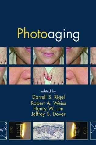
The results of UV publicity and getting older have gotten an more and more serious factor in scientific perform. solar publicity and different resources of UV radiation play an immense function within the early visual appeal of good and coarse wrinkles, roughness, laxity, abnormal pigmentation, and roughness of the surface. This medical consultant is the 1st of its type to explain the big array of remedies on hand for the minimization and service of the results of photodamage—prompting clinicians to tailor healing regimens to person elements of getting older and stability the hazards and advantages of every remedy opposed to sufferer expectancies.
Read or Download Photoaging PDF
Similar medicine books
Panagiotis A. Tsonis's Anatomy of Gene Regulation: A Three-dimensional Structural PDF
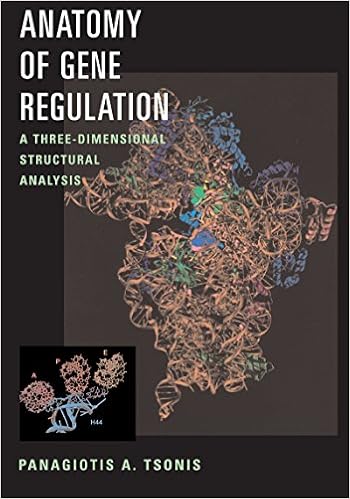
Not uncomplicated line drawings on a web page, molecular constructions can now be considered in full-figured glory, frequently in colour or even with interactive probabilities. Anatomy of Gene legislation is the 1st e-book to provide the components and methods of gene legislation on the third-dimensional point. vibrant constructions of nucleic acids and their significant other proteins are published in full-color, three-d shape.

- Stem Cells and Regenerative Medicine: Patents and Clinical Trials (Stem Cells - Laboratory and Clinical Research Series)
- Pocket Medicine: The Massachusetts General Hospital Handbook of Internal Medicine (4th Edition)
- Impressions for Implant Dentistry make easy
- Essentials of Orthopedic Surgery
- Development of a rational scale to assess the harm of drugs of potential misuse
- Hemostasis and Thrombosis: Practical Guidelines in Clinical Management
Extra info for Photoaging
Example text
Clinical and Histological Changes 47 Figure 11 Lentigo maligna. Atypical melanocytes are present in a lentiginous pattern at the dermoepidermal junction, and extend into the basal layer of hair follicles. A lentigo malignant melanoma develops in an in situ lentigo maligna. The clinical presentation is initially the same as lentigo maligna in situ, plus the development of gray areas (indicating focal regression) or blue areas (indicating dermal pigment, melanocytes, or melanin), or papules and nodules.
B* colorimetric plane (13). 14 The values proposed for the angles of skin categories boundaries are: Very light skin Ͼ 55Њ Ͼ Light skin Ͼ 44Њ Ͼ Intermediate skin Ͼ 28Њ Ͼ Mat/Tan skin Ͼ10Њ Figure 6 illustrates the relationship between skin color typing defined by the ITAЊ values and MED values determined on the back of volunteers 24 hr after exposure to solar simulator (xenon arc lamp complying with Colipa recommendations) (unpublished results, 2003). As shown on this figure, the MED values decrease when ITAЊ values increase.
The most frequent repair mechanism used by the cells is called excision repair. Altered bases or nucleotides are removed and replaced by new, undamaged elements. The enzymes involved in this repair system are glycosylases, endonucleases, and polymerases. Photoreactivation, the other repair mechanism, involves only one enzyme, photolyase, which reverses the damage using the energy of light. Photolyase seems to be absent or nonfunctional in humans. However, it has recently been shown that topical application of this enzyme on human skin repairs the CPDs induced by UV radiation (23).
Photoaging
by Joseph
4.3