
By Swennen G.R.J. (ed.), Schutyser F.A.C. (ed.)
This richly illustrated color atlas and guide offers orthodontists, maxillofacial and plastic craniofacial surgeons, genetic dysmorphologists and scientific anthropologists with exhaustive info on all facets of third-dimensional cephalometric research of demanding and tender tissues. The e-book deals sensible, straight forward ''step-by-step'' counsel for either clinicians and researchers attracted to three-D review of the top and face.
Read Online or Download Three-Dimensional Cephalometry: A Color Atlas and Manual PDF
Best medicine books
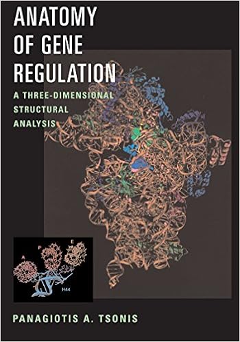
Anatomy of Gene Regulation: A Three-dimensional Structural - download pdf or read online
Now not uncomplicated line drawings on a web page, molecular constructions can now be seen in full-figured glory, usually in colour or even with interactive percentages. Anatomy of Gene rules is the 1st booklet to offer the elements and tactics of gene legislation on the three-d point. vibrant buildings of nucleic acids and their better half proteins are published in full-color, 3-dimensional shape.

- Ion Beam Treatment of Polymers. Application aspects from medicine to space
- Spondylotherapy; physio-therapy of the spine based on a study of clinical physiology
- The Rosiecrucian secrets: Their excellent method of making medicines of metals also their lawes and mysteries
- What They Didn't Tell You At Medical School
- Perspectives in receptor research: proceedings of the 10th Camerino-Noordwijkerhout Symposium, Camerino, Italy, 10-14 September 1996
Extra info for Three-Dimensional Cephalometry: A Color Atlas and Manual
Example text
Cadaver skull). b The skull of a 6-year-old child: dorsal view. 1 Axial CT Slices b Fig. 19. ). ) Fig. 20. 3-D hard-tissue surface representation shows the position of orbitomeatal orientated axial slices 1–8 (Figs. ) 37 CHAPTER 2 Basic Craniofacial Anatomical Outlines Axial CT – Slice 1 Fig. 21. ). 2 Multiplanar CT Anatomy of the Skull CHAPTER 2 Axial CT – Slice 2 Fig. 22. 1 Frontal bone; 2 Frontal sinus; 3 Orbital roof; 4 Optic canal; 5 Anterior cranial fossa; 6 Sphenoid bone; 7 Sphenosquamosal suture; 8 Temporal bone 39 CHAPTER 2 Basic Craniofacial Anatomical Outlines Axial CT – Slice 3 Fig.
A The skull base of a 6-year-old child: exocranial view (cadaver skull). b The skull base of a 6-year-old child: exocranial view (3-D CT, cadaver skull) a b Fig. 17. a The skull of a 6-year-old child. Superior view (cadaver skull). b The skull of a 6-year-old child. 1 3-D CT Anatomy of the Skull a CHAPTER 2 b Fig. 18. a The skull of a 6-year-old child: dorsal view. (cadaver skull). b The skull of a 6-year-old child: dorsal view. 1 Axial CT Slices b Fig. 19. ). ) Fig. 20. 3-D hard-tissue surface representation shows the position of orbitomeatal orientated axial slices 1–8 (Figs.
B The skull base with the mandible removed: exocranial view (3-D CT, adult cadaver skull) 21 CHAPTER 2 Basic Craniofacial Anatomical Outlines Skull – Superior View (Calvaria) Fig. 5. a Superior view of the skull (calvaria) (adult cadaver skull). 1 3-D CT Anatomy of the Skull CHAPTER 2 Fig. 5. b Superior view of the skull (calvaria) (3-D CT, adult cadaver skull) 23 CHAPTER 2 Basic Craniofacial Anatomical Outlines Calvaria – Interior View Fig. 6. a Interior view of the calvaria (adult cadaver skull).
Three-Dimensional Cephalometry: A Color Atlas and Manual by Swennen G.R.J. (ed.), Schutyser F.A.C. (ed.)
by Charles
4.0